02 / Hepatic · CT
LiverX
BETAHepatic fat and tissue density, read from a scan you already took.
01 · It comes with the cardiac scan
No second protocol.
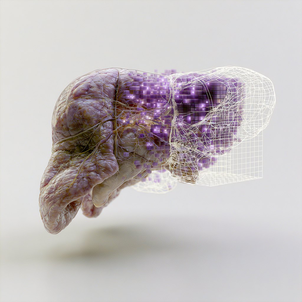
A non-contrast cardiac CT already contains the liver. LiverX reads it out of the volume HeartX has ingested — no extra acquisition, no extra dose, no extra visit. If your protocol already scans the chest, the hepatic readout is a configuration, not a study amendment.
Measured within the cardiac field of view — not a substitute for a dedicated abdominal CT.
02 · What it measures
Three hepatic readouts from a cardiac volume.
Hepatic fat fraction
The proportion of liver voxels in the fat attenuation range, computed from the volume.
Parenchymal density
Mean and low-percentile attenuation across the tissue.
Lobar morphometry
Volume and shape of the liver within the field of view.
Each measurement is anchored to published thresholds and ships with the method that produced it. The methodology →
03 · Why it matters now
Liver fat is a trial endpoint.
Metabolic and MASH programmes — SGLT2, GLP-1 — are measuring hepatic fat already, usually with a separate imaging visit. If a cardiac CT is in the protocol, the measurement is already in the data.
04 · Status
Where this module is.
Extraction, per-patient values, cohort view and export, on every cardiac study that contains the liver.
Calibrating published thresholds for this field of view, and comparison against dedicated abdominal CT on the same patients.
The loop wrote this module’s specification and flagged its own limits before any code existed. How the loop works →
Tell us what your protocol already scans.
If a cardiac CT is already in the schedule, the liver is already in the volume.